Following the previous interpretation of the cleavage stage assessment rulesInterpretation of the New Istanbul Consensus (Part 2): Key Content of Cleavage Stage (Days 1-3) Embryo Assessment, this article will focus on the blastocyst stage assessment content in the new Istanbul Consensus. With the maturity of blastocyst culture technology, blastocyst transfer has become a routine clinical practice (only 2% of ART centers do not perform blastocyst transfer at all), and the proportion of blastocyst cryopreservation is significantly higher than that of cleavage-stage embryos (more than 50% of centers only freeze embryos at the blastocyst stage). The new consensus is based on the most widely used Gardner scoring system in clinical practice, supplemented and revised with a large amount of clinical data, clarifying the core criteria for blastocyst assessment, while refining special morphologies for which existing evidence is insufficient to confirm relevance to quality, avoiding over-exclusion.
1. Scoring System: New Grade D Blastocyst to Distinguish Between Non-Viable and Low-Viable
The Gardner scoring system assesses blastocyst quality through three dimensions: expansion stage, inner cell mass (ICM) grade, and trophectoderm (TE) grade, and it remains the most widely used scoring scheme in clinical practice (63% of respondents use this scheme). The new consensus adds a grade D blastocyst to the original three grades A/B/C (representing excellent, good, and fair respectively). Grade D blastocysts are specifically used to mark non-viable blastocysts with degenerative features (such as massive cell degeneration) or without a clearly identifiable ICM, so as to avoid confusion with grade C blastocysts (grade C blastocysts have low viability but still have certain clinical potential). The new consensus clearly states that grade D blastocysts are not recommended for clinical use, while grade C blastocysts can still be used cautiously after comprehensively considering the specific conditions of patients.

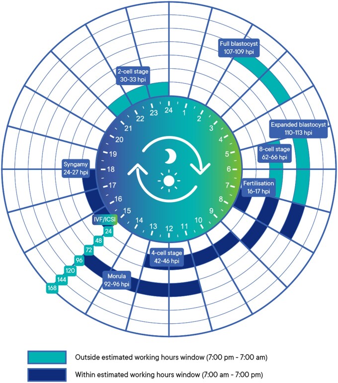
2. Blastocyst Formation Days: All Days 4-7 Are Included in the Assessment, Clarifying Prognostic Differences Among Days
The number of days of blastocyst formation directly reflects the developmental speed of the embryo and is closely related to its viability. Based on multicenter TLT data and clinical outcome analysis, the new consensus includes blastocysts formed from day 4 to day 7 in the assessment range, emphasizing the number of days after insemination as the core judgment basis and combining the laboratory’s own culture system to record the assessment time.
- Day 4 Blastocysts: Refers to embryos that have formed a blastocoel when observed statically on the 4th day after insemination, with a low clinical incidence (<5%). The consensus shows that if such blastocysts reach the expansion stage (Gardner stage 3 and above), the existing data does not clarify whether their implantation rate after frozen transfer is significantly different from that of day 5 blastocysts. However, it is necessary to be alert to the risk of aneuploidy associated with their excessively fast development. Therefore, it is recommended to confirm the chromosome ploidy with PGT-A technology before transfer, especially for elderly patients (>35 years old).
- Day 5 Blastocysts: This is the most ideal time for blastocyst formation and is the first choice for fresh transfer and cryopreservation. Consensus data shows that at 108 hpi (hours post-insemination), 47%-52% of embryos develop into complete blastocysts, and they have a significant advantage in euploidy rate compared with day 6 and day 7 blastocysts. In addition, the fresh transfer live birth rate (about 24%-30%) and frozen transfer live birth rate (about 22%-28%) of day 5 blastocysts are the highest among all days, and the pregnancy loss rate after transfer is also relatively low, so they are suitable as a priority.
- Day 6 Blastocysts: Refers to blastocysts formed on the 6th day after insemination. Their live birth rate (about 17%-22% for fresh transfer and 15%-20% for frozen transfer) is lower than that of day 5 blastocysts, but they still have clinical value. The consensus suggests that if there are no available blastocysts on day 5, blastocysts that reach Gardner 3BB and above on day 6 can be used for transfer in patients with a small number of embryos (the original paper does not mention the specific number of oocytes retrieved); and frozen transfer can partially make up for the impact of developmental delay (the gap in live birth rate between day 6 frozen blastocysts and day 5 is reduced to 3%-5%).
- Day 7 Blastocysts: Account for 5%-10% of all available blastocysts. The original paper does not specify their aneuploidy rate and implantation rate. Although the consensus does not provide specific data, it clarifies their overall quality status. For patients with older maternal age (>38 years old), very few oocytes retrieved (≤3), or repeated implantation failure, if day 7 blastocysts reach Gardner 3BB and above and PGT-A indicates euploidy, they can still be used as one of the transfer options, but patients must be fully informed that the risk of pregnancy loss is relatively high (about 25%-35%).
3. Key Prognostic Indicators: TE Grade Is the Most Important, Multi-Dimensional Comprehensive Assessment
Multivariate analysis (including factors such as expansion stage, ICM grade, TE grade, and blastocyst formation days) shows that the indicators clearly related to blastocyst implantation potential are ranked by importance as follows, and laboratories should pay priority attention during assessment:
- Trophectoderm (TE) Grade: It is the strongest predictor of live birth. TE cells are responsible for placental formation after embryo implantation, and their quality directly affects the success rate of implantation. Blastocysts with TE grade A (cells arranged tightly, uniformly, with clear boundaries) or B (cells arranged relatively tightly, with a small number of gaps) have a significantly higher live birth rate than those with grade C (cells arranged loosely, with many gaps).
- Expansion Stage: The higher the degree of expansion, the higher the probability of live birth. The Gardner scoring system divides the expansion stage into 6 stages. The viability of blastocysts in stages 3-6 is significantly higher than that in stages 1-2, and they should be given priority in clinical selection.
- Inner Cell Mass (ICM) Grade: The impact is still unclear, and its impact on outcomes is weaker than that of TE. Existing studies show that there is no significant difference in the live birth rate between blastocysts with ICM grade A or B; only when the ICM grade is C may it increase the risk of pregnancy loss, but there are still successful live birth cases. Therefore, embryos cannot be excluded solely because the ICM grade is C, and a comprehensive judgment should be made based on the TE grade and the number of days of the blastocyst.
4. Special Morphologies and Clinical Selection: Comprehensive Judgment Based on Evidence, Avoid Over-Screening
In addition to core special morphologies such as double ICM and low-grade blastocysts, the following special situations need to be carefully evaluated based on existing evidence. They cannot be directly regarded as irrelevant factors, nor should they be over-focused or excluded:
- Residual Cells/Fragments Under the Zona Pellucida: A small amount of degenerated cells or fragments may remain under the zona pellucida of some blastocysts. At present, it is only clear that the degree of fragmentation in cleavage-stage embryos is related to developmental potential. In studies on residual fragments under the zona pellucida at the blastocyst stage, no association with reduced implantation rate or live birth rate has been found, nor has it been classified as an abnormal morphology to be excluded. During clinical evaluation, priority should be given to the overall expansion degree of the blastocyst, the grades of ICM and trophectoderm (TE), and a comprehensive judgment should be made based on the overall quality of the embryo. The clinical value of the embryo cannot be denied solely based on the presence of a small amount of residue under the zona pellucida.
- Special Hatching Patterns: Atypical hatching patterns such as peanut-shaped, figure-eight-shaped, or only a small amount of TE hatching may occur during blastocyst hatching. The current core of blastocyst hatching assessment lies in the expansion stage (such as stages 3-6 in the Gardner score) and overall structural integrity, such as whether TE protrudes normally from the zona pellucida and whether the blastocyst completely detaches from the zona pellucida. It does not involve the association between specific types of hatching morphology and embryo quality or implantation potential, and there is no evidence that atypical hatching patterns will affect the subsequent developmental outcome of the embryo. In clinical practice, it is only necessary to confirm that the overall structure of the blastocyst is intact, and there is no need to directly exclude it due to special hatching morphology.
- Impact of Spontaneous Collapse on Euploid Embryos: About a quarter of blastocysts will experience spontaneous collapse, manifested as temporary disappearance and then re-expansion of the blastocoel. Studies have found that blastocysts with spontaneous collapse are more likely to be aneuploid, and the collapse of aneuploid blastocysts is often associated with a lower implantation rate; however, for blastocysts with confirmed euploid chromosomes, a history of collapse does not affect their implantation potential. If preimplantation aneuploidy testing (PGT-A) is not performed, the value of the blastocyst can be judged by observing whether it can re-expand. If the morphology is normal after expansion, its clinical application possibility can still be retained, and it cannot be completely excluded solely based on the occurrence of collapse.
- Cytoplasmic Strings: Cytoplasmic strings are dynamic structures connecting TE and ICM cells, participating in intercellular communication, and can be observed in 55%-85% of expanded blastocysts for transfer. Such structures are positively correlated with the implantation rate of blastocysts and can be used as a supplementary basis for blastocyst sorting. However, it should be noted that the assessment of cytoplasmic strings is somewhat subjective, and the consistency between different observers and the same observer at different times is only at a general to moderate level. Moreover, there is insufficient evidence to show that it can be used as an independent core standard for judging embryo quality. In clinical practice, cytoplasmic strings can be included in the auxiliary assessment dimension, and a comprehensive judgment should be made based on core indicators such as blastocyst expansion stage and TE/ICM grade. The choice of embryo cannot be determined solely based on the presence or absence of cytoplasmic strings.
With this, through the systematic interpretation of three articles, the core practice guidelines of the 2025 new Istanbul Consensus on embryonic development timeline, pronuclear stage assessment, cleavage stage assessment, and blastocyst stage assessment have been fully presented, providing comprehensive and accurate clinical references for embryologists and reproductive medicine practitioners, and helping assisted reproductive technology to better serve clinical practice under a standardized and normalized framework.